February 21, 2025 | Deborah Kotz
Utilizing a novel diagnostic test for peripheral blood immunophenotyping, researchers were able to identify the specific sources of inflammation triggering the novel skin disease and reverse symptoms with targeted treatments

In a recent paper published in the Nature journal Scientific Reports, researchers from the University of Maryland School of Medicine described a new skin disease in a male patient with erythroderma, causing 80 percent of his skin to be covered with red, exfoliating skin lesions that itched and burned. After undergoing months of treatment with traditional therapies for erythroderma, which included the steroid prednisone, anti-itch creams, and immunosuppressive drugs, the patient experienced little relief.
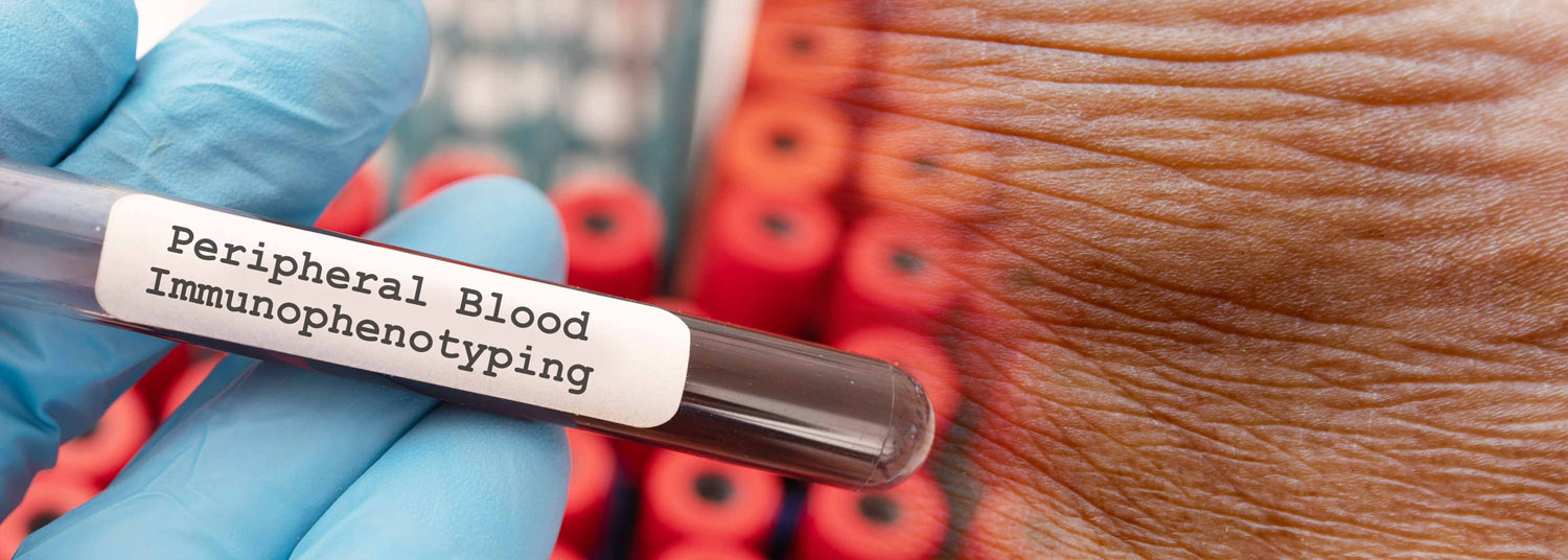
“We isolated individual circulating blood cells and created a new blood test using flow cytometry to identify specific cytokine signatures,” said study corresponding author Shawn Kwatra, MD, the Joseph W. Burnett Endowed Professor and Chair of Dermatology at UMSOM and Chief of Service Dermatology at the University of Maryland Medical Center (UMMC). The authors received a patent for this new method, involving “peripheral blood flow cytometry-based immunophenotyping enabled us to identify a novel form of a severe and potentially life-threatening skin disease.”

To determine which of the immune system’s components were driving the inflammatory disease, Dr. Kwatra and his team used a new flow cytometry platform technique, for which they received a patent, to immunophenotype skin diseases. They found that two of these cytokines, called interleukin-13 and interleukin-17, were at increased levels in this patient compared to healthy controls as well as when compared to patients with other known causes of erythroderma. Subsequently, targeted treatment with biologic inhibitors of IL-13 and IL-17 reversed the patient’s disease.

When the patient was treated with a dual therapy of two monoclonal antibodies, dupilumab and secukinumab, his symptoms dramatically decreased and eventually resolved, essentially curing him of his erythroderma. The authors also identified the cell sources of these pathological cytokines and monitored the decline in immunopathogenic (disease-causing) cell numbers, and the decline of interleukin-13, and interleukin-17 levels in the patient's blood throughout the treatment course.
“We created a new diagnostic test to discover a previously undescribed skin disease and initiate appropriate treatment. We are now exploring developing our diagnostic test to a range of other inflammatory skin,” said Dr. Kwatra.
The study was funded by the National Institutes of Health. Co-authors from Duke University School of Medicine, George Washington University School of Medicine, and Johns Hopkins University School of Medicine, also contributed to the research.
“This research represents a promising first step towards the development of sophisticated diagnostic tools that employ immunophenotyping to pinpoint the causes of non-specific inflammatory conditions. Patients with these conditions urgently need access to precision-based therapies to help them better manage their symptoms and lead productive lives,” said Mark T. Gladwin, MD, who is the John Z. and Akiko K. Bowers Distinguished Professor and Dean of University of Maryland School of Medicine.
Contact
Deborah Kotz
dkotz@som.umaryland.edu