Core Facilities
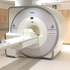
3 Tesla Biograph mMR Scanner
Description: State-of-the-art body scanner for simultaneous PET-MRI, 60cm open bore
Manufacturer: Siemens Healthineers
Click here for manufacturer's website
Details
Please select a section for more details:
Capabilities
- Precise alignment of MR and PET through simultaneous acquisition
- Single frame of reference
- Minimal motion artifacts
- Brain functional MRI
- Cardiac function
Specifications MRI
- 60cm open bore
- Parallel imaging with 64 receive channels
- 20 channel head RF coil
- MQ Gradients (45 mT/m @ 200 T/m/s)
- Software version VE11P
- Broadband capability, including 11C
Specifications PET
- LSO Crystals
- 60cm open bore
- NEMA 4.4mm transverse resolution
- Axial FOV 258mm
Contact
Rao Gullapalli, PhD
Phone: 410-706-2694
rgullapalli@som.umaryland.edu
Jiachen Zhuo, PhD
Phone: 410-706-2697
jzhuo@som.umaryland.edu
Thomas Ernst, PhD
Phone: 410-706-1045
ternst@som.umaryland.edu
Peripheral equipment
- Rear-projection system for visual presentation
- MR-compatible earphones
- Handheld button response boxes
- Mock scanner
- Camera-based KinecCor motion tracking system
Template for NIH "Facilities and Other Resources"
This is a template for the MR-PET scanner, which can be used in the "Facilities and Other Resources" page of NIH applications. Before using this template, please contact your collaborator to ensure the information is correct and up to date.
"The combined PET-MRI scanner is a state-of-the-art Siemens 3.0 Tesla mMR Biograph system with zero helium boil-off. The system has a high-performance transmit and receive system and the applications are available for head-to-toe imaging along with high-performance gradients for a combined system. Maximum gradient amplitude is 45 mT/m for each direction, i.e. 78 mT/m vector summation gradient performance, and the max slew rate is 200 T/m/s. Minimal rise time of 224 microseconds is possible from 0-45mT/m. The system is also equipped with a broad-band multinuclear multi-nuclear imaging capability to facilitate imaging of F-19, Na-23, C-13 etc.
The PET system uses LSO crystals with a ring diameter of 656mm with a total of 28,672 crystal elements. The scanner facilitates an axial field of view of 258mm and 127 image planes, and includes advanced PET attenuation correction using 5-compartment model including bones from head to thigh."

