2023 Archive
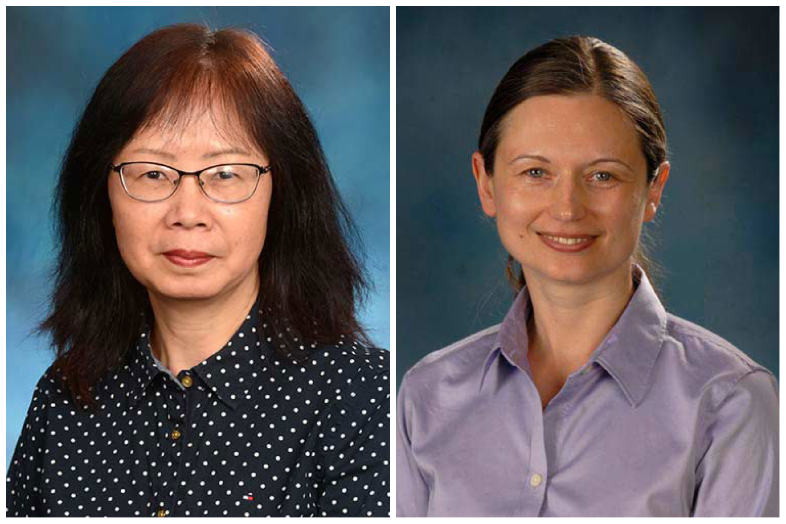
Congratulations to Drs. Wu and Lipinski on their recent publications in PNAS and Science Advance
February 20, 2023

Traumatic brain injury accelerates age-related lipofuscin formation in microglia
Aged microglia tend to accumulate lipofuscin (LF), which is considered to be an intralysosomal “garbage” or waste material, primarily composed of proteins and lipid like material. An autofluorescent (AF) in aging microglia named LF reflects a pathological state leading to microglial senility and immune dysfunction. LF is associated with phagocytosis and inflammatory neurodegeneration that can be further accelerated by a traumatic brain injury (TBI), according to new research in mice and human published on March 8th in leading international journal, Science Advances.
Dr. Junfang Wu, Professor of Anesthesiology and Shock, Trauma and Anesthesiology Research (STAR) Center in Maryland's School of Medicine and focused on microglia in the brain and spinal cord, is the lead author of the research.
LF and AF in microglia are dramatically increased with normal aging, however, the functional relationship between lipid-laden microglia in age-related neurological dysfunction and head injury has yet to be described.
Dr. Rodney M. Ritzel, who is now an assistant professor at UTHealth Houston and collaborated with experts at the University of Maryland School of Medicine, report the first detailed immunophenotypic analysis of naturally occurring, age-associated AF microglia in brain of both mice and human. This subpopulation of microglia in older mice adopt a unique dysfunctional phenotype defined by increases in phagocytosis, oxidative stress levels, lysosomal content and autophagy markers, lipid and iron accumulation, metabolic alterations, pro-inflammatory cytokine production, and senescent-like features. The team mechanistically demonstrated that the AF phenotype is more pronounced by chronic brain injury-induced oxidative stress and APOE4 genotype at middle and old age. The latter is associated with increased risk of developing late-onset Alzheimer's disease (AD). Finally, the researcher inhibited a particular receptor microglia need to survive. The inhibition can eliminate the AF microglial signature, reduce inflammatory burden, improve motor and cognitive performance, and prophylactically protect the brain against severe head impact.
Journal Reference:
- Rodney M. Ritzel, Yun Li, Yun Jiao, Zhuofan Lei, Sarah J. Doran, Junyun He, Rami A. Shahror, Rebecca J. Henry, Romeesa Khan, Chunfeng Tan, Shaolin Liu, Bogdan A. Stoica, Alan I. Faden, Gregory Szeto, David J. Loane, Junfang Wu. Brain injury accelerates the onset of a reversible age-related microglial phenotype associated with inflammatory neurodegeneration. Science Advances, 2023; 9 (10) DOI: 10.1126/sciadv.add1101
Autophagy protein ULK1 interacts with and regulates SARM1 during axonal injury
Marta Lipinski, PhD, Associate Professor, Department of Anesthesiology and Shock, Trauma and Anesthesiology Research (STAR) Center, authored of "Autophagy protein ULK1 interacts with and regulates SARM1 during axonal injury," which was published in PNAs on November 14, 2022.
Axonal degeneration is observed and plays an important role in acute CNS injury and in neurodegenerative diseases. Previous studies found that autophagy, a cellular catabolic pathway generally thought to be neuroprotective, can promote axonal degeneration through a mechanism that remains poorly understood. Our studies of axonal degeneration in a mouse model of spinal cord injury (SCI) demonstrate a functional and physical interaction between upstream regulator of autophagy ULK1 and the essential axonal degeneration mediator SARM1. Our findings suggest a direct regulatory crosstalk between autophagy and axonal degeneration pathways, which is mediated through ULK1-SARM1 interaction and contributes to the ability of SARM1 to accumulate in injured axons.
Journal Reference:
- Choi, H. M. C., Li, Y., Suraj, D., Hsia, R.-C., Sarkar, C., Wu, J., & Lipinski, M. M. (2022). Autophagy protein ULK1 interacts with and regulates SARM1 during axonal injury. Proceedings of the National Academy of Sciences, 119(47). https://doi.org/10.1073/pnas.2203824119
Contact
Department of Anesthesiology
(410) 328-6120 (phone)
(410) 328-5531 (fax)
newsletter@som.umaryland.edu
